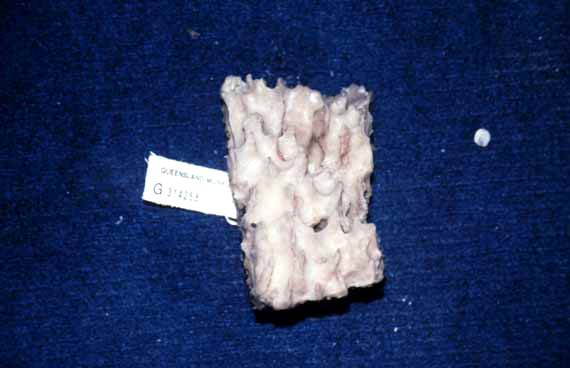
Morphological description (show/hide)
| massive | | beige | | not apparent | | soft, flaccid, interior feels gritty due to coring of large fibres by foreign matter | | conulose, conules large, ranging from evenly distributed to large and sweeping in some specimens, conules with distinctive ends, pointed or ridged, but all specimens have at least some pointed conules, interior reticulation easily observed macroscopically | | The choanosomal skeleton consists of several components. There is a basal layer of spongin, 60-240 ï¾µm thick, lying on dead coral or on other sponges (sometimes with foreign sponge spicules protruding into the basal skeleton), and with an erect basal layer of smooth choanosomal principal styles and echinating acanthostyles standing perpendicular to the substrate, which together form a leptoclathriid structure. Intermingled with, or overlaying the erect basal skeleton is a regularly renieroid reticulation of acanthostrongyles in pauci- or multispicular (vaguely ascending) and uni- or paucispicular (irregularly transverse) tracts. Meshes are triangular to rectangular, 60-150 ï¾µm maximum diameter, without any obvious spongin fibre component, lined by abundant but virtually unpigmented interfibril spongin, and with circular to oval choanocyte chambers 63-95 ï¾µm in diameter. The mesohyl matrix of the basal skeleton (i.e. point of attachment to the substrate) and peripheral skeleton is more heavily pigmented than spongin in the choanosomal region, and microscleres appear to be more abundant near the surface. At the major nodes of the renieroid skeleton are echinating acanthostyles, occurring singly or in plumose brushes, and these may form irregularly plumose, usually discontinuous ascending tracts. In addition to the renieroid skeleton is a subisodictyal extra-fibre skeleton of choanosomal and subectosomal megascleres. Those spicules originate and extend from the erect leptoclathriid basal skeleton, often ascending all the way to the surface. Extra-fibre tracts become increasingly plumose and dendritic towards the periphery, sometimes forming a subisodictyal reticulation, but those megascleres only form clearly continuous tracts in thicker sections, whereas in thinly encrusting specimens extra-fibre spicules may be more irregularly dispersed throughout the choanosomal skeleton. | | The ectosomal skeleton is microscopically hispid, with the points of smooth choanosomal principal styles protruding through the surface. Detritus is usually absent from the surface, although thinly encrusting specimens may contain isolated arenaceous particles. The projecting principal megascleres are surrounded at their bases by plumose brushes of mostly small ectosomal auxiliary subtylostyles, although larger subectosomal megascleres may also participate in ectosomal brushes. The subectosomal region is structurally variable. In thinly encrusting sections the peripheral skeleton is not clearly delineated from the choanosomal skeleton, containing only thick tangential to paratangential tracts of larger subectosomal auxiliary subtylostyles, with tracts measuring up to 140 ï¾µm thick in some sections. In thicker bulbous sections the subectosomal region is relatively cavernous, containing numerous plumose, stellate brushes of choanosomal and subectosomal megascleres, and that region is clearly differentiated from the renieroid choanosomal skeleton. Subectosomal megascleres also commonly occur in the deeper choanosomal skeleton, together with smooth choanosomal principal styles, and those spicules may form vaguely ascending, multispicular, extra-fibre tracts, 25-65 ï¾µm in diameter, or they may be irregularly dispersed throughout the skeleton. | | nil | | nil |
|

