Morphological description (show/hide)
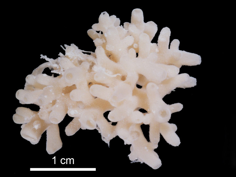
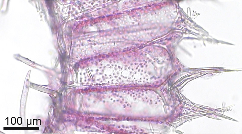
| With a cormus of dichotomous to polychotomous branching tubes and a specimen size of 3_3 cm, torn in two pieces. Tubes are cylindrical and have a diameter of 2-3 mm, with the central tubes slightly larger. | | white | | beige-white | | One prominent osculum is present apical on each tube, with its diameter slightly smaller than the tube itself. A small oscular rim is common. The osculae are at least partially enclosed by a thin membrane. | | Soft, easily torn. | | Minutely tesselated. | | Inarticulated choanosomal skeleton. Perpendicular longer paired actines of subcortical pseudosagittal triactines support the distal part of the choanosomal skeleton, with the longer unpaired actines of subatrial sagittal triactines forming the proximal part. No spicule tracts are present, but occasionally tri/tetractines are added to the choanoskeleton, with their paired actines/basal triradiate system tangentially arranged in the middle to proximal third of the choanocyte chambers. Their unpaired/apical actines point towards the ectosome The subatrial skeleton is formed by shorter paired actines of sagittal subatrial triactines. The atrial skeleton consists of sagittal tetractines with longer and slightly curved apical actines, which protrude into the atrium (figure10b), and sagittal triactines, with their basal system tangentially arranged. | | Bundles (tufts) of microdiactines form the minutely tesselated external surface over the distal ends of the radially arranged choanocyte chambers. The base of these diactine- bundles is formed by one (or more) t-shaped sagittal triactines, with their unpaired actine in the centre of the bundle of microdiactines, perpendicular to the external surface of the tube. Paired actines are tangentially arranged at the top of the choanocyte chambers and are close to the unpaired- and shorter paired-actine of subectosomal pseudosagittal triactines. Over the top of the inhalant canals (located between the radial choanocyte chambers) tangentially arranged triactines are found. The oscular rim is formed by tangential ectosomal t-shaped triactines, with occasional diactines perpendicular to the surface. | | Ectosomal, parasagittal to sagittal triactines: paired actines 40-(53.8)-77 um, SD 9.6 um], unpaired actine 40-(54)-88 um, SD 8.7 um ], both x 4-(6.9)-9 um (SD 1.3 um). Choanosomal pseudosagittal triactines: longer and curved paired actine: 73-(108.2)-130 um, SD 14.5 um, shorter paired actine 67-(95)-132 um, (SD 17.6 um), and unpaired actine 36-(68.8)-85 um, (SD 9.2 um), all 3-(8.5)-12 um (SD 1.5 um) thick. Occasionally triactines and rarely tetractines were found in the skeleton of the choanocyte chambers, but these do not form an articulated choanoskeleton. It is not clear whether these represent subatrial spicules (having the same dimensions) dislocated during section preparation, or true choanosomal spicules as they were sparse in all section preparations. Subatrial sagittal triactines: unpaired actines 97-(155.8)-246 um long, (SD 35.5 um). Their tangential shorter paired actines form the subatrial skeleton, 53-(81.8)-170 um long (SD 25.9um), 3-(8.5)-12um maximum thickness (SD 2.1 um). Plough- like sagittal tetractines form the atrial skeleton (figure 11f ), with their free and mostly bent apical actine protruding into the atrium. The apical actine is thickened and curved in the opposite direction of the unpaired actine of the basal triradiate system. The basal triradiate system is mostly parasagittal, with more-or-less regular angles but a longer unpaired actine. Atrial tetractines with a mostly parasagittal basal triradiate system [unpaired actines of the basal triradiate system 86-(157.2)-262 um (SD 47.4 um),paired actines slightly shorter, both 6-(10.6)-15 um (SD 2.6 um) thick, apical actines: 102-(167.9)-366 mm (SD 62 mm) and slightly thicker. Tangential atrial triactines with dimensions similar to ectosomal triactines. | | Ectosomal microdiactines are mostly straight but sometimes slightly sinuous and tapering proximally to a more-or-less sharp end. Some have a distal lance-head, which starts at about three-quarters from the proximal end, but may be longer, whereas others do not show a lance- head but only a slight thickening from where the spicule tapers towards the ends:54-(84.7)-119_3-(4.4)-6 um (SD 17.1_1.1um). |
|


