Morphological description (show/hide)
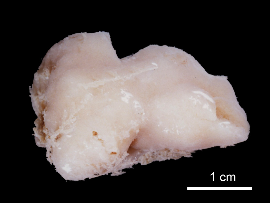
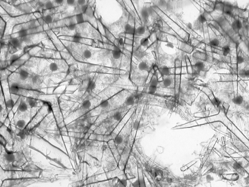
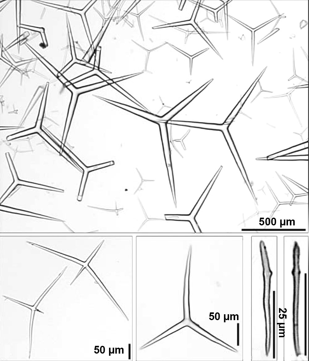
| Small massive, elongated tubes, clustered together with a lobate/knobby surface. With a specimen size of 3x2x2 cm. | | white | | pale beige | | Four naked oscules apical on the massive tubes, with a diameter of 1-4 mm, and a small lip. | | Firm and harsh to touch, but not hispid | | Convoluted, knobby. Partially overgrown by hydrozoans, partly covered with sediment. | | Clearly differentiated from the cortex, consisting of an irregular articulated meshwork of large regular triactines, arranged without apparent order throughout the sponge body. Some actines of the more superficial choanosomal triactines (with respect to the cortex) protrude through the cortex. Choanosomal triactines are densely packed, choanocyte chambers scattered between. Smaller triactines are rare, apparently representing developmental stages of choanosomal triactines as opposed to cortical ones. The small incurrent water canals appear to have no special skeleton, but are difficult to discern due to the strongly developed skeleton. A special skeleton, consisting of peculiar sagittal tetractines and rare triactines (with similar peculiar shape as the tetractines but lacking apical actines) characterizes the excurrent canals. Up to 10 tetractines (with rarely intercalated triactines) are densely packed above each other with their basal triradiate system tangential to the walls of canals. Smaller apical actines of tetractines extend into the canals. Microdiactines, indistinguishable in size and shape from the cortical ones, are present in the walls of the excurrent canals. They appear to be concentrated around the openings of the smaller excurrent canals extending into the terminal canals (leading to the oscules), where they are embedded perpendicular to the walls of the canals in the small membrane (ÔlipÕ) above these openings. | | A distinct thin cortex is present, 100-200 um thick. The distal (external) part of the cortex is supported by more-or-less tangentially arranged small, slightly sagittal triactines, remarkably different in size from those found in the choanosome. Microdiactines are dispersed irregularly throughout the cortex, and also sometimes arranged perpendicularly in bundles at the external surface. Actines of larger perpendicular choanosomal triactines sometimes protrude through the cortex. |
|


