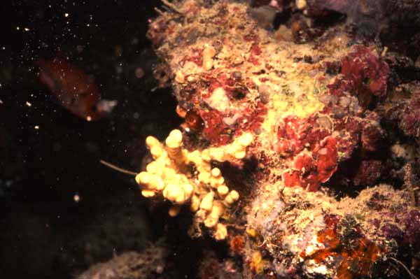
Morphological description (show/hide)
| Thickly encrusting clump, approximately 22 x 10 cm, composed of lobate-bulbs scattered over the dead coral surface (Plate 1A). | | Live colouration is yellow-brown alive (Munse | | Oscula: Large oscula are raised above the surface of the sponge on conical membranous pedicels, and these appear to be confined to the upper external surfaces of lobes (Plate 1B). Minute ostia (<0.5 mm diameter) are numerous and evenly distributed over the upper (exposed) surface of lobes, with a few slightly larger examples situated within shallow exhalant drainage canals (Plate 1B). | | Sponge consistency is relatively soft, easily compressible and easily torn. | | The surface is macroscopically even in situ, but minutely hispid, even shaggy in places in preserved material, produced by the terminal choanosomal spicule brushes. Scattered over lobes are evenly rounded bumps and bulbs, and the sponge appears insubstantial due to the numerous small oscula scattered over the surface. Most bulbous lobes are excavated by one or more shallow drainage canals. membranous pedicels on which oscula are raised, seen in live material (Plate 1b), collapse upon preservation. | | The choanosomal skeleton is plumo-reticulate. There is no trace of axial compression of the skeleton, but in the centre of each lobate bulb the spicule tracts form regular or irregularly reticulate, triangular or square isodictyal meshes, up to 180 ᄉm in diameter, bounded on all sides by uni-, pauci- or multispicular tracts of rhabdostyles (Fig. 7). Spongin fibres are poorly invested in type A spongin, but spicule tracts also have heavy deposits of spongin B surrounding them, and these deposits are particularly heavy in the axis of the skeleton. In the extra-axial region of the choanosomal skeleton, towards the periphery, spicule tracts become clearly separated into primary ascending multispicular tracts, with 3-6 spicules in each row, and secondary transverse unispicular components, one or two spicules in length. Transverse spicules and spicule tracts diverge from the ascending fibres at angles of 30-90﾿, and these could be interpreted as echinating spicules (e.g. Hallmann 1917). However, most of these transverse secondary spicule tracts in the extra-axial skeleton interconnect with the adjacent primary ascending fibres, producing a vaguely isodictyal reticulation, whereas at the periphery they are clearly plumose, so the term echinating may be misleading. Choanocyte chambers are oval, 150-260 ᄉm in diameter. The mesohyl matrix is very heavily invested with dark brown type B spongin, which contains numerous microscleres. | | The ectosomal skeleton is membranous, without any specialized spiculation, but a prominent feature of this region is the protruding spicule brushes from the primary choanosomal tracts (Fig. 6). These tracts of rhabdostyles diverge near the surface, becoming increasingly plumose, and spicule brushes may extend for up to 400 ᄉm from the surface. Spicule brushes of the peripheral skeleton are loosely bound together with heavy granular type B spongin. This ectosomal spongin contains numerous irregularly scattered microstyles, but these also occur in equally heavy concentrations elsewhere in the skeleton. | | Choanosomal rhabdostyles are relatively robust, thick, with rhabdose bases bent at between 35-70﾿ from the shaft, evenly rounded, unspined, and never contort, occasionally styles are seen without rhabdose bases, but these are rare. The apex varies from fusiform sharply pointed in smaller spicules to hastate-pointed in larger examples, and the shaft usually contains a sparse scattering of small spines in the distal two-thirds of the spicule (Fig. 9): 178-(235.1)-283 x 3-(14.0)-22 ᄉm. | | Microstyles are relatively long, thin, sometimes straight but usually with a flexuous bend near the middle, with a slightly subtylote base, tapering to sharp raphidiform points. Microstyles have prominent microspination over their shafts and rounded bases, like other members of the genus, but these spines appear only as a slight roughening of the surface under light microscopy, whereas higher magnification clearly shows individual spines (Fig. 10): 53-(82.4)-96 x 0.8-(1.5)-2.0 ᄉm.Toxas are sparsely microspined (Fig. 12), small, thin, ranging from v-shaped to forms with a gentle central curvature and reflexed arms: 18-(39.4)-72 x 0.4-(0.9)-1.2 ᄉm.Sigmas are small, thin, contort, usually with a central curl, but occasionally they are regularly c-shaped. Sigmas are shown to be smooth under light microscopy, but higher magnification reveals prominent microspination, as for microstyles and toxas (Fig. 11): 6-(12.2)-15 x 0.5-(0.8)-1.2 ᄉm. |
|

